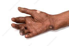
LEPROSY (HANSEN’S DISEASE):
Leprosy is also called as Hansen’s disease since the causative organism first isolated by Dr. Gerhard armauer Hansen in 1874.It is a chronic non fatal infectious disease caused by slow growing bacteria called Mycobacterium leprae. It affects mainly the cooler parts of the body such as skin, eyes, nose, mouth, respiratory tract, earlobes and peripheral nerves. Temperature for growth is 30C.In leprosy the earliest and the main involvement is of skin and nerves.In India, leprosy is the major public health problem affecting millions of people. AS lepra bacillus does not grow on artificial media and cannot be transmitted to all animals, it is difficult to culture this organism and study the effect of drug. The disease is termed as a chronic granulomatous disease, it is similar to that of tuberculosis, because it produces inflammatory nodules that is granulomas on the skin and nerves over time.
• It is caused by slow growing Rod-shaped, acid fast bacteria called as
Mycobacterium leprae or Mycobacterium lepromatous.It is an obligate
intracellular pathogens that usually lives in macrophages.
• The organisms that is Mycobacterium lepromatous Or laprae in tissues appear as a compact rounded masses (globi) or are may arranged in parallel fashion like similar to cigarettes-in- pack.
• M. leprae can be demonstrated by using tissue sections, in split skin smears by
splitting the skin, scrapings from cut edges of dermis, and in nasal smears by the
following techniques:
1. Acid-fast (Ziehl-Neelsen) staining:The staining procedure is similar as for
demonstration of M. tuberculosis but can be decolourised by lower concentration
(5%) of sulphuric acid (less acid-fast)
2. Fite-Faraco staining procedure is a modification of Z.N. procedure and is considered better for more adequate staining of tissue sections.
3. Gomori methenamine silver (GMS) staining can also be used.
4. Molecular methods by PCR.
5. IgM antibodies to PGL-1 antigen seen in 95% cases of lepromatous leprosy but only in 60% cases of tuberculoid leprosy.
INCIDENCE:
The disease is endemic in areas with hot and moist climates and also in poor tropical countries. According to the WHO, 8 countries-India, China, Nepal, Brazil, Indonesia, Myanmar (Burma), Madagascar and Nigeria, together constitute about 80% of leprosy cases, of which India accounts for one-third of all registered leprosy cases globally. In India, the disease are more commonly seen in regions like Tamil Nadu, Bihar, Pondicherry, Andhra Pradesh, Orissa, West Bengal and Assam. Very few rare cases are now seen in Europe and the United States.
Jopling’s Handbook of Leprosy Hardcover – 30 July 2020. Buy Now
LEPROSY SYMPTOMS:
Symptoms of leprosy is mainly affect the skin, mucus membrane (cooler Or moist area inside the body) and the nerve.
Skin manifestations
• Presence of discoloured patches on the skin is observed. The patches are usually flat and faded (the colour is lighter than the skin present around)
• Growth of nodules on the skin
• Skin stiffness
• Dry skin
• Thickening of skin
• Ulcers may present in the soles of the feet, which are usually painless
• Lumps on the face
• Swelling
• Loss of eyelashes or eyebrows are observed
Neurological manifestations
• Numbness on affected areas of the skin
• May cause muscle weakness or paralysis of hand and feet
• Nerve enlargement
• Vision problems that may leads to blindness
• Injuries such as burns may go unnoticed
Symptoms cause by the leprosy on the mucous membrane includes
• Stuffy nose
• Nose Bleeding
Untreated or advanced leprosy
• Paralysis and crippling of hands and feet
• Blindness
• Loss of sensation
• Nose disfigurement
• Painful nerves
• Loss of eyebrows
• Burning sensation of skin
• Shortening of toes
• Redness of affected area
TYPES OF LEPROSY:
Leprosy can be categorized into three types includes,
1. Lepromatous leprosy: This leprosy is also called as a Multibacillary(MB). It is
characterized by weaker humoral antibody mediated immune system. In this type
of leprosy the host lacks resistance to bacteria and more severe type of leprosy.
Forms more or numerous skin lesions. Shows significant bacterial growth. This is
highly infectious to others
The following are common features characterise lepromatous polar leprosy
i) In the dermis, there may be proliferation of macrophages with foamy change,
particularly around the blood vessels, nerves and dermal appendages. The
foamy macrophages are called as ‘lepra cells’ or Virchow cells.
ii) The lepra cells are heavily filled with acid-fast bacilli which can demonstrated
with help of AFB staining.
iii) The dermal infiltrate of lepra cells ,it does not encroach upon the basal layer
of epidermis and which is separated from epidermis by a formation of sub
epidermal uninvolved clear zone.
iv) The epidermis overlying the lesions are thinned and flat and may even
ulcerate.
2. Tuberculoid leprosy: This type of leprosy is also called as Paucibacillary (PB). It is characterized by the hypo or hyper pigmented skin macules that leads to the loss of sensation. It may be characterized by a stronger cell mediated immune response that is nothing but a cytotoxic T cells. In this type of leprosy the host is highly resistant to bacteria and less severe form of leprosy. Forms less skin lesions when compared to Multibacillary. Low infective to others.
The polar tuberculoid form involves the following histological features
i) The dermal lesions shows the formation of granulomas resembling hard
tubercles which is composed of epithelioid cells, Langhans’ giant cells and also
the peripheral mantle of lymphocytes.
ii) Lesions of tuberculoid leprosy have predilection for dermal nerves which may
be destroyed and infiltrated by epithelioid cells and also the lymphocytes.
iii)The granulomatous infiltrate destroys the basal layer of epidermis that is
there is no formation of clear zone.The presence of lepra bacilli are few and
are maybe seen in destroyed nerves
3. Borderline leprosy: This type of leprosy is also called as diamorphous. It has both the characteristics features of both Multibacillary leprosy and Paucibacillary
leprosy. The characteristics features of the three forms of borderline leprosy are as under as follows
i) Borderline tuberculoid (BT) form shows epithelioid cells and plentiful
lymphocytes. There is a formation of narrow and clear sub epidermal zone.
Lepra bacilli are scanty and are found in nerves.
MODE OF TRANSMISSION:
Leprosy is a slow communicable disease
• Human to human transmission is the primary source of infection, three other
species can carry and rarely transfer Mycobacterium leprae to human include
chimpanzee, money, Mangabey and nine-banded armadillos
• Direct contact with untreated leprosy patients who shed numerous bacilli from
damaged skin, nasal secretions, mucous membrane of the mouth and also from hair follicles.
• Droplet infection
• Materno foetal transmission across the placenta
• Transmission from milk of leprosy affected mother to infant during breastfeeding
• Airborne transmission through inhalation of the Bacilli
• Tattooing needles
• Contact with soil or fomites
• Transmission through insect vectors like mosquito, bed bugs etc
What is the incubation period? Incubation period between first exposure and appearance of signs and symptoms of the disease varies between 2-20 years (average about 3 years)
RISK FACTORS:
• Close contact with leprosy patient
• Genetics
• Age
• Immunosuppression or immunocompromised individual are more prone to leprosy infection
COMPLICATIONS OF LEPROSY:
• Blindness or glaucoma
• Progressive disfiguration of the face
• Erectile dysfunction that may cause the infertility in men
• Kidney failure
• Crippling of hands
• Hair loss
• Muscle weakness
• Permanent damage to the inside of the nose which leads to the bleeding of nose
• Permanent nerve damage
• Loss of sensation on skin
PREVENTION OF LEPROSY:
The prevention of leprosy lies between the early diagnosis and treatment of those
individuals suspected or diagnosed as having leprosy, thereby preventing further
transmission of the disease to other people.
• Public education and community awareness plays an crucial role to encourage
individuals with leprosy and their families to undergo evaluation and treatment
with Multi drug therapy
• Household contacts of patients with leprosy should be closely monitored for the
development Or any of leprosy signs and symptoms
• A study demonstrated that prophylaxis with a single dose of rifampicin was 57%
effective in preventing leprosy for the first two years in individuals who have close
contact with newly diagnosed patients with leprosy. There is currently no widely
used standard for using medications for the prevention of leprosy
• Currently, there is no single commercial vaccine that confers complete immunity
against leprosy in all individuals.
• Several vaccines including the BCG vaccine, provide variable levels of protection against leprosy in certain populations.
DIAGNOSIS OF LEPROSY:
• History: which includes patients bio data (age, sex) and family history of leprosy
• Clinical examination:which include the inspection of the surface of the body that is skin and testing for loss of sensation and paralysis
• Bacteriological examination:which includes skin smears and nasal smears
• Foot pad culture:used for detecting drug resistance and also for detecting the
viability of Bacilli during the treatment
• Immunological test:this is a test for cell mediated immunity.
LEPROMIN TEST:
It is not a diagnostic test but this test is used for classifying leprosy on the basis of
immune response. On Intra- dermal injection of lepromin(an antigenic extract of
Mycobacterium leprae)gives delayed hypersensitivity reaction in patients with
tuberculoid leprosy:
• An early positive reaction appearing as an indurated area in 24-48 hours is called Fernandez reaction.
• A delayed granulomatous lesion appearing after 3-4 weeks is called Mitsuda
reaction. Patients of lepromatous leprosy shows negative by the lepromin test.The test indicates that cell-mediated immunity is greatly suppressed in case of lepromatous leprosy while patients of tuberculoid leprosy show good immune response in lepromin test. Delayed type of hypersensitivity is conferred with helper T cells. The granulomas of tuberculoid leprosy have sufficient helper T cells but only fewer number of helper T cells are observed in case of lepromatous leprosy.
LEPROSY TREATMENT:
Major goals of treatment are
• Early detection of patients
• Appropriate treatment
• Adequate care to be taken for the prevention of disabilities and rehabilitation
MDT(Multi drug therapy) Treatment includes taking antibiotics, Dapsone and rifampin Steroid medication are used to reduce the pain and acute inflammation in patient with leprosy


